Pages
Health Care News
Categories
- Asthma education
- Autism
- Canadian Health&Care Mall
- Cardiac function
- Critical Care Units
- Follicle
- Health
- health care medical transport
- health care programs
- Health&Care Professionals
- Hemoptysis
- Hormone
- Isoforms
- Nitroglycerin Patches
- Profile of interleukin-10
- Progesterone
- Pulmonary Function
- Sertoli Cells
- Theophylline
- Tracheoesophageal Fistula
Canadian HealthCare News
Structural and Functional Modifications of Sertoli Cells: DISCUSSION(9)
Considering the abnormality seen in the Sertoli cell cytoplasm, the question arises whether it is possible that such cells degenerate with time, thus compromising the seminiferous epithelium. In fact, at no time did we observe pyk-nosis of Sertoli cell nuclei, degeneration of their organelles, or evidence of apoptotic figures in the seminiferous epithelium. In addition, the basal Sertoli cell cytoplasm and junctional complexes between adjacent Sertoli cells appeared intact. Furthermore, the absence of an increased number of macrophages or neutrophils in the interstitial space of the testis of FORKO mice suggests that there is no leakage of substances from the epithelium as a result of the swelling of the Sertoli cell cytoplasm or complete breakdown of the blood/testis barrier.
(more…)
Structural and Functional Modifications of Sertoli Cells: DISCUSSION(8)
 In addition, the onset of tubular fluid production at 20-35 days coincides with a prepubertal rise in FSH. Based on these observations and our findings of a dilated Sertoli cell cytoplasm in FORKO mice, it seems likely that FSH-R regulates water balance in the Sertoli cells. Recent findings reported by Haywood et al. using the hpg mouse model lend support to this hypothesis. These mice lack gonadotropins, and constitutively express a mutated FSH-R, showing increased levels of tubular fluid secretion resulting from overactive FSH-R signaling.
(more…)
Structural and Functional Modifications of Sertoli Cells: DISCUSSION(7)
These types of studies have not been addressed in the various Sertoli cells of a given seminiferous tubule at a given stage of the cycle. Nevertheless, the reason why Sertoli cells have evolved such complex mechanisms of protein regulation is unclear, but the data suggest that the testis has some capacity to maintain itself in part under adverse conditions.
(more…)
Structural and Functional Modifications of Sertoli Cells: DISCUSSION(6)
 As Sertoli cells normally stop dividing by Day 21, we did not count the number of Sertoli cells in adult FORKO mice in this study; there was no evidence of mitosis of Sertoli cell nuclei at any ages examined in the present investigation.
The finding that some tubules in a cross-section appear normal while others show varying degrees of altered epithelium is apparently not unique to the FORKO testis and has also been reported for other knockout mouse models. While the underlying reasons for such a phenomenon are still not fully understood, several speculations can be advanced.
(more…)
Structural and Functional Modifications of Sertoli Cells: DISCUSSION(5)
In addition, the tubules that were affected usually displayed a semilunar disruption, i.e., only one half of the tubule was affected in a cross-section. Affected tubules were found at all stages of the cycle of the seminiferous epithelium, suggesting that this phenomenon is not stage specific. The quantitative data indicated very clearly that, while focal areas of Sertoli cell cytoplasm appear swollen, this effect does not extend globally to the entire tubule itself, which actually is reduced by 16%, 35%, and 30% in profile area at 3, 6, and 12 mo, respectively in the FORKO mice relative to the wild-type mice.
(more…)
Structural and Functional Modifications of Sertoli Cells: DISCUSSION(4)
 As Sertoli cells are difficult to visualize in standard fixation and staining preparations, with only their nuclei being prominent, we utilized an antiprosaposin antibody, a specific marker of Sertoli cells, to stain these cells. Prosaposin has been localized to Sertoli cells, irrespective of the stage of the cycle, and in conjunction with immunocytochemis-try, the antibody readily stains these cells. In this way, we were able to note that Sertoli cells in FORKO mice showed a radial stellate-shaped distribution around each tubule in a manner similar to that seen in wild-type mice.
(more…)
Structural and Functional Modifications of Sertoli Cells: DISCUSSION(3)
Second, concurrent perfusions of mice from many other knockout models studied in our laboratory by identical procedures never resulted in dilated spaces of the type seen in the FORKO mice, indicating that these spaces are not related to the fixative or the method used to perfuse FORKO mice. Third, dilated cytoplasmic areas in different generations of germ cells were never observed in the FOR-KO mice. The dilations were exclusively restricted to cytoplasm of Sertoli cells, suggestive of something specific about the relationship of the knockout to these particular cells.
(more…)
Structural and Functional Modifications of Sertoli Cells: DISCUSSION(2)
 In contrast, in estrogen receptor alpha knockout mice, the epithelium of the efferent ducts is affected, resulting in fluid accumulation in the seminiferous tubular lumen and disruption of the epithelium. More apically, the dilated spaces surrounded the heads of elongating spermatids (Fig. 11). Being membrane bound, such spaces corresponded to the cytoplasm of the apical processes of Sertoli cells that are known to envelop the heads of spermatids in various mammals at different stages of the cycle.
(more…)
Structural and Functional Modifications of Sertoli Cells: DISCUSSION(1)
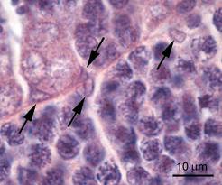
The most striking aspect of the present study was the finding of large irregularly shaped translucent spaces situated between germ cells in the testis of FORKO mice. For a variety of reasons, it was concluded that these spaces within the epithelium were territories of the Sertoli cell cytoplasm that were highly dilated and filled with fluid. In appropriate planes of section, it was noted that the basal cytoplasm of Sertoli cells at times appeared intact, showing a nucleus and organelles embedded in a finely flocculent ground substance (Fig. 11). These intact areas were confluent with the highly dilated space that extended toward the lumen.
(more…)
Structural and Functional Modifications of Sertoli Cells: RESULTS(6)
 Quantitative Analyses
Quantitative measurements indicated that the mean cross-sectional profile area of seminiferous tubules in FOR-KO mice was significantly lower compared with wild-type mice at all ages examined (Fig. 9 and Table 1). In addition, both groups showed significant changes in mean profile areas of the tubules as the age of the animals increased (Fig. 9 and Table 1). The mean profile area of tubules in wild-type mice rose by about 5700 ^m2; per tubule from 3 mo and 6 mo and then increased by an additional 14 300 ^m2 per tubule between 6 mo and 12 mo of age (Fig. 9).
(more…)
