Pages
Health Care News
Categories
- Asthma education
- Autism
- Canadian Health&Care Mall
- Cardiac function
- Critical Care Units
- Follicle
- Health
- health care medical transport
- health care programs
- Health&Care Professionals
- Hemoptysis
- Hormone
- Isoforms
- Nitroglycerin Patches
- Profile of interleukin-10
- Progesterone
- Pulmonary Function
- Sertoli Cells
- Theophylline
- Tracheoesophageal Fistula
 |
Canadian Health&Care; MallVisit the most reliable Canadian Health&Care; Mall offering a wide choice of drugs for any medical emergency you may have, from male health to infections and obesity! Making sure you always spend less money is among our top priorities! |
Disturbed Expression of Sox9 in Pre-Sertoli Cells: RESULTS(1)
Timing and Distribution of Pre-Sertoli Cells in B6. Ytir Fetal Gonads
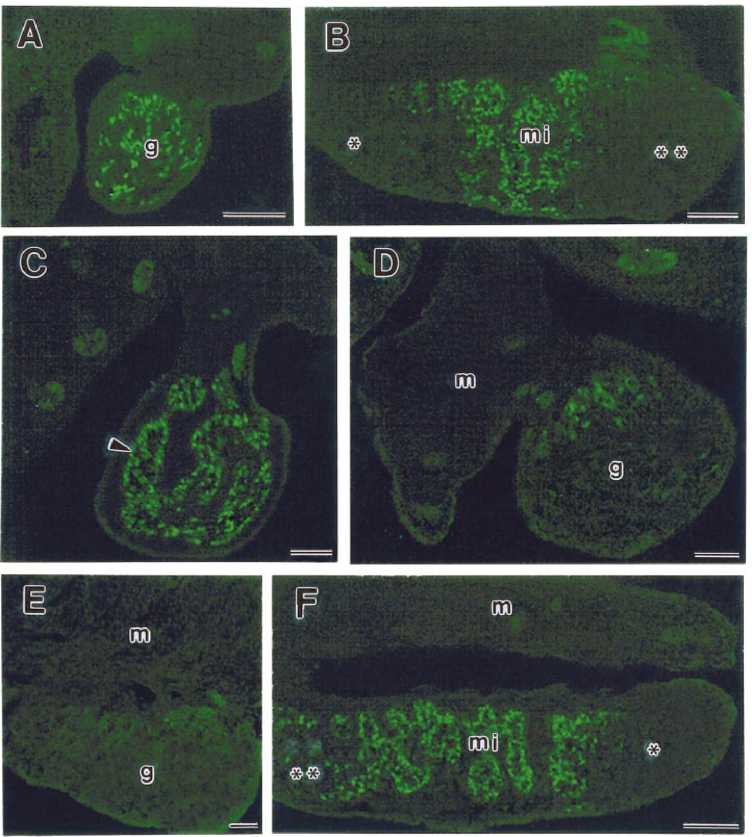
Pre-Sertoli cells were identified by immunofluorescent staining with an antibody against Sox9 protein and were observed in all 12 B6.Ytir gonads at 11.5 and 12.5 dpc (Table 1). Longitudinal and cross-serial sections of urogenital ridges revealed a distribution pattern of pre-Sertoli cells within the still undifferentiated genital ridges. Cross serial sections revealed that pre-Sertoli cells were located among the innermost cells of the thickening genital ridges and rarely formed part of the coelomic epithelium (Fig. 1A). Longitudinal sections showed that pre-Sertoli cells were located in the medial region, whereas they were absent from both the cranial and caudal regions of the genital ridges (Fig. 1B).
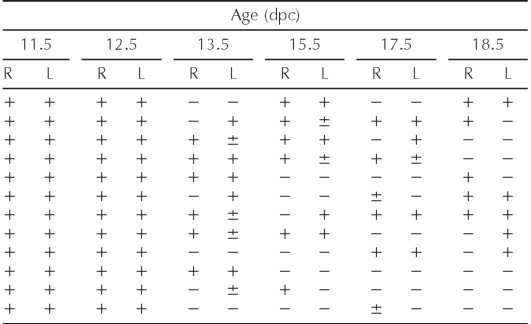
Interestingly, whereas at 13.5 dpc Sox9-positive cells were present in both gonads of six out of 12 serially sectioned B6.Ytir embryos, they were considerably diminished in one gonad of three of these embryos. In three other embryos both gonads lacked Sox9-positive cells, and in the other three embryos Sox9-positive cells were absent in one of the two gonads (Table 1).
 FIG. 1. Indirect immunofluorescent staining for Sox9 in frozen sections of B6.Ytir embryos at two developmental stages. A) and B) Urogenital ridges at 12.5 dpc. A) Cross-section of the middle region showing green-fluorescent Sox9-positive cells located in the core of the genital ridge (g). B) Longitudinal section showing Sox9-pos-itive cells located in the medial region (mi). Notice the absence in the cranial and caudal regions. C-F) gonads at 13.5 dpc. C) Cross section of the middle region showing that Sox9-positive cells are forming seminiferous cords (arrowheads). D) and E) Cross- and tangential sections of urogenital complexes showing few Sox9-positive cells in the gonad. These cells appear located in the dorsal side of the gonad (g) near the mesonephros (m). F) Longitudinal section showing organized seminiferous cords in the middle region (mi) of the gonad. Whereas in the cranial region several scattered Sox9-positive cells appear, in the caudal region they are absent. Bar = 100 ^m.
TABLE 1. SOX9 epression in serially sectioned gonads of B6.Ytir embryos.*
 Right (R) and left (L) gonads. N = 12 B6. Ytir embryos at each age. +, ±, or — = abundant, few, or absent SOX9-expressing cells, respectively.
 FIG. 1. Indirect immunofluorescent staining for Sox9 in frozen sections of B6.Ytir embryos at two developmental stages. A) and B) Urogenital ridges at 12.5 dpc. A) Cross-section of the middle region showing green-fluorescent Sox9-positive cells located in the core of the genital ridge (g). B) Longitudinal section showing Sox9-pos-itive cells located in the medial region (mi). Notice the absence in the cranial and caudal regions. C-F) gonads at 13.5 dpc. C) Cross section of the middle region showing that Sox9-positive cells are forming seminiferous cords (arrowheads). D) and E) Cross- and tangential sections of urogenital complexes showing few Sox9-positive cells in the gonad. These cells appear located in the dorsal side of the gonad (g) near the mesonephros (m). F) Longitudinal section showing organized seminiferous cords in the middle region (mi) of the gonad. Whereas in the cranial region several scattered Sox9-positive cells appear, in the caudal region they are absent. Bar = 100 ^m.
TABLE 1. SOX9 epression in serially sectioned gonads of B6.Ytir embryos.*
 Right (R) and left (L) gonads. N = 12 B6. Ytir embryos at each age. +, ±, or — = abundant, few, or absent SOX9-expressing cells, respectively.
Tags: developmental biology gene regulation ovary Sertoli cells
