Pages
Health Care News
Categories
- Asthma education
- Autism
- Canadian Health&Care Mall
- Cardiac function
- Critical Care Units
- Follicle
- Health
- health care medical transport
- health care programs
- Health&Care Professionals
- Hemoptysis
- Hormone
- Isoforms
- Nitroglycerin Patches
- Profile of interleukin-10
- Progesterone
- Pulmonary Function
- Sertoli Cells
- Theophylline
- Tracheoesophageal Fistula
 |
Canadian Health&Care; MallVisit the most reliable Canadian Health&Care; Mall offering a wide choice of drugs for any medical emergency you may have, from male health to infections and obesity! Making sure you always spend less money is among our top priorities! |
Structural and Functional Modifications of Sertoli Cells: RESULTS(6)
 Quantitative Analyses
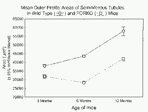
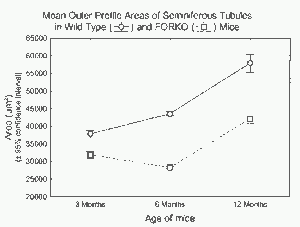
Quantitative measurements indicated that the mean cross-sectional profile area of seminiferous tubules in FOR-KO mice was significantly lower compared with wild-type mice at all ages examined (Fig. 9 and Table 1). In addition, both groups showed significant changes in mean profile areas of the tubules as the age of the animals increased (Fig. 9 and Table 1). The mean profile area of tubules in wild-type mice rose by about 5700 ^m2; per tubule from 3 mo and 6 mo and then increased by an additional 14 300 ^m2 per tubule between 6 mo and 12 mo of age (Fig. 9).
The mean profile area of tubules in FORKO mice, however, decreased by about 3500 ^m2 per tubule from 3 mo and 6 mo and then increased by 13 900 ^m2 per tubule, roughly the same increment detected for wild-type mice between 6 mo and 12 mo of age (Fig. 9). These within-group changes in cross-sectional profile areas of the seminiferous tubules were significant across all ages (Table 1).
Western Blots
Western blots of testicular extracts from wild-type and FORKO mice probed with the anti-ABP antibody showed the presence of reactive bands, as expected, near 75 kDa. The reactive bands appeared more intense in the extracts from the wild-type mice compared with the FORKO mice at equivalent total protein loads per lane (Fig. 10; density data). Another set of reactive bands near 30 kDa was also observed in these blots and were caused by anti-GST im-munoreactivity simultaneously present in the anti-ABP antibody (the antigen used to elicit the antibody was a recombinant GST-fusion protein) (data not shown). This antigen expressed in the testicular Leydig cells served as the control for equal protein loading in different lanes (data not shown).
 FIG. 9. Mean cross-sectional profile area of seminiferous tubules in wild-type (circles, solid line) and FORKO (squares, dashed line) mice at different ages. The mean profile area of tubules in FORKO mice is significantly less than (P < 0.05) the mean profile area of tubules in wild-type mice at all ages. The mean profile area of the tubules also increases markedly between 3 mo and 12 mo of age, and in the same ratio from 6 mo to 12 mo of age, in both groups.
 FIG. 11. Schematic drawing of a portion of the seminiferous epithelium of a wild-type (upper) and FORKO (lower) mouse at 3 mo of age as seen in the electron microscope. In wild-type mice (upper panel), there is a close relationship between Sertoli cells and adjacent spherical and elongating germ cells. The cytoplasm of the Sertoli cell is compact, containing numerous organelles embedded in a finely flocculent ground substance. The thin, attenuated apical Sertoli cell processes (Sp) extend between the elongating spermatids seen near the lumen and contain various organelles. The ectoplasmic specializations (ES, also seen in inset) adhere to the heads of elongating spermatids and are composed of filaments (f) and flattened ER cisternae. The blood testis barrier (BTB) is intact and seen in the basal region of the epithelium. In FORKO mice (lower panel), the Sertoli cell cytoplasm is greatly distended and there is a lack of structural epithelial integrity in areas where abnormalities prevail. Also prominent are large, dilated apical Sertoli cell processes, which encompass elongating spermatids. In such areas, there is an absence of the finely flocculent ground substance, and few organelles are evident amid membranous whorls (MW) and profiles of varying shapes and sizes. Ectoplasmic specializations (ES, also seen in inset), while apparent, show fewer filaments (f) and occasional dilated ER cisternae. In the basal region, the blood testis barrier (BTB) is present and apparently intact. G, Golgi apparatus; m, mitochondria; Ly, lysosomes; In, invagination; A, acrosome; N, nucleus of Sertoli cell; Spct, spermatocyte; Spga, spermatogonia; LSptd, late spermatid; ESptd, elongating spermatid. Illustration by Haitham Badran.
TABLE 1. Descriptive statistics (A) and univariate ANOVA tests (B) on log10 trasformed data.
| Summary results for outer profile areas (^m2) | |||
| Group | Age (no. | ) | Mean ± SD (num. obs.) |
| Wild type | 3 | 37 822 ± 8815 | |
| 6 | 43 580 ± 4543 | ||
| 12 | 57 918 ± 17 089 | ||
| FORKO | 3 | 31 863 ± 7244 | |
| 6 | 28 289 ± 2737 | ||
| 12 | 42 179 ± 10 477 | ||
| Two-factor univariate ANOVA tests of significance for outer profile areas | |||
| Degrees of | |||
| Effect* | SS | freedom | MS F P |
| Treatment | 5.4218 | 1 | 5.4218 652 0.0000 |
| Age | 5.3993 | 2 | 2.6996 325 0.0000 |
| Treatment X age | 0.6651 | 2 | 0.3325 40 0.0000 |
| * Treatment (wild type of FORKO); age (3, 6, or 12 mo.). | |||
Tags: follicle-stimulating hormone receptor male reproductive tract Sertoli cells sperm testis
